


October 1, 2025
The Hidden Cost of Retakes
For many dental teams, retakes have become such a normal part of daily practice that they are accepted as routine. One quick retake may not seem like a big deal—until you realize how often it’s happening. Each retake interrupts clinical flow, adds minutes to chair time and increases patient exposure. Multiply that by several patients a day and suddenly the cumulative cost of “just one more exposure” becomes significant.
Every retake forces clinicians and assistants to stop what they are doing, reposition the patient, re-explain the process and often reassure an uneasy patient. What could have been a smooth, seamless procedure becomes a disruption. For practices that pride themselves on efficiency and patient comfort, this can create unnecessary frustration.
Research shows that retakes are among the most frequent sources of inefficiency in imaging. In fact, a 2025 study found that 37.2% of intraoral periapical radiographs required repetition, most often due to positioning errors and cone misalignment. These errors not only take time but also directly impact diagnostic quality, as repeated exposures may still fail to produce the sharp, consistent images clinicians rely on for treatment planning.
Beyond workflow, retakes affect patient experience. Patients may become anxious or frustrated when asked to sit still for multiple exposures and keep a hard sensor in their mouths for longer than necessary. They may also worry about radiation exposure, even if clinicians reassure them that doses are minimal. According to the American Dental Association (ADA), radiation exposure from dental X-rays is low, but the principle of ALARA—“As Low As Reasonably Achievable”—remains a standard of care. Every unnecessary exposure is one that could have been avoided with the right equipment and processes.
How Retakes Disrupt Workflow
From a business perspective, time is one of the most valuable resources in dentistry. When an X-ray has to be retaken, it does more than consume a few extra seconds. It creates a ripple effect that can reduce productivity across the practice.
First, it delays the clinician’s ability to move forward with diagnosis or treatment. If an image is unclear, the appointment stalls until a diagnostic-quality image can be captured. This delay may extend chair time and reduce the number of patients a clinician can see in a day.
Second, it interrupts team workflow. Assistants who might otherwise be preparing an operatory for turnover or helping another patient are pulled back into repeating a process that should have been completed on the first try. Over the course of a day, these small interruptions add up, leading to bottlenecks in patient flow.
Finally, it can impact patient satisfaction. Patients value efficiency and often equate it with competence. When they perceive that the dental team is struggling to capture an image, confidence may erode. This matters not only for the current appointment but for long-term trust and case acceptance.
A workflow study published in the Journal of Dental Hygiene noted that imaging errors and retakes are among the most common workflow disruptors in dental practices. Even a modest reduction in retake frequency can free up significant chair time, enabling clinicians to focus on what matters most—patient care.

The Role of Equipment Design
Not all retakes are due to operator error. Often, they are the result of equipment limitations. One of the most common culprits is tubehead and/or arm drift. When an X-ray unit’s arm fails to stay in place, even slight movement can blur an image or shift the beam just enough to miss the intended target. The result is a retake.
Some mounted, hardwired intraoral X-ray units are designed to reduce this problem. Unlike portable units with weaker support structures, a properly engineered, drift-free arm holds steady exactly where the clinician positions it. This stability reduces the likelihood of image distortion and makes it easier for assistants and clinicians to work confidently, knowing the unit will remain in place.
Reliability also plays a role. A unit that is always ready—powered, mounted and in position—removes the variability of setup. There is no need to track down equipment, wait for a charge or wonder whether the unit was left in another operatory. It is always where you left it, always ready for the next patient. This predictability is a subtle but powerful way to improve efficiency.
From Efficiency to Clinical Confidence
Reducing retakes isn’t only about saving time. It’s also about improving clinical outcomes. Each clear, diagnostic-quality image supports a more confident diagnosis. Clinicians can detect dental caries, evaluate bone health and plan restorative or surgical treatment with greater accuracy when images are sharp and consistent.
High-frequency direct current (DC) technology, as found in many modern units, produces more consistent images with less radiation than older alternating current (AC) units (see Dentalcare.com CE course). Features like a smaller focal spot also enhance image sharpness, providing clinicians with the detail they need to identify subtle changes in tooth or bone structure.
The result is often a virtuous cycle: fewer retakes, faster workflow, sharper images and more accurate diagnoses—all of which help improve patient trust. When patients see clear images and understand the rationale behind treatment recommendations, they are more likely to accept proposed care.

The Patient Perspective
For patients, intraoral imaging is often one of the least comfortable parts of a dental visit. Holding film or digital sensors in the mouth can trigger gag reflexes or discomfort. Being asked to repeat the process only compounds the experience.
Patients may also perceive repeated imaging as unnecessary exposure to radiation, regardless of how minimal the dose. While studies have shown that digital imaging can reduce exposure significantly—by as much as 80% compared to traditional film—patients are increasingly aware of radiation safety and may ask questions. Being able to reduce retakes and capture high-quality images the first time helps build trust.
Beyond exposure, patients value efficiency. A smooth appointment with minimal interruptions reinforces the perception that their provider is skilled and well-equipped. Conversely, repeated delays can erode that confidence. For practices competing in an environment where patient expectations are higher than ever, these details matter.
What to Look for in an Intraoral X-Ray Unit
Choosing the right intraoral X-ray unit requires balancing technical performance with practical workflow needs. Key considerations include:
- Image quality: A smaller focal spot and DC technology can improve sharpness and reduce radiation.
- Reliability: A drift-free arm that stays in place helps ensure consistent capture.
- Workflow integration: A mounted, hardwired unit can save setup time and is ready when needed.
- Usability: Intuitive controls and ergonomic design can help reduce the learning curve for new staff and minimize positioning errors.
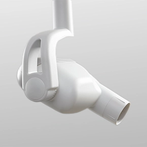
The Midmark Preva™ Intraoral X-Ray was engineered with these factors in mind so clinicians can capture high-quality images the first time, every time. Key benefits of the Midmark Preva include:
- Drift-free, sag-free design that holds position, helping reduce retakes, minimize interruptions and maintain diagnostic confidence
- Consistent, high-quality images with a 0.4-mm focal spot and drift-free design
- Always-ready convenience with mounted, hardwired units that are both reliable and predictable—always where you left them, always ready for the next patient
- Ease of use for the team with intuitive controls, selectable exposure settings and a compact, ergonomic design—helping simplify training and daily operation

Conclusion: Breaking the Retake Routine
Retakes are more than minor inconveniences. They disrupt workflow, extend chair time, increase radiation exposure and create unnecessary frustration for both patients and teammates. For clinicians, they represent lost time that could be spent on diagnosis, treatment and building patient relationships.
Modern intraoral X-ray units designed for stability, reliability and ease of use can dramatically reduce retakes. The result is often greater efficiency, sharper images and better patient care.
For practices looking to improve both productivity and patient experience, investing in the right X-ray unit is more than an equipment upgrade—it’s a way to eliminate one of the most persistent sources of frustration in dentistry.
Ready to reduce the likelihood of retakes and streamline workflow? Discover how the can help your practice capture the image right the first time.