


The ECG Is Only as Good as the Acquisition: Why ECG Accuracy Starts with Technique

By:
Clinical Solutions Advisor, Midmark


We See It

Rethinking the Exam Room - AIA Credit


The right procedure room equipment can make a crucial difference in both patient outcomes and caregiver safety. Particularly for patients...
Explore the 631 Procedure Chair


By:
Clinical Solutions Advisor, Midmark
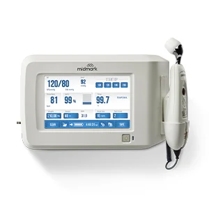
A 12-lead electrocardiogram (ECG /EKG) is one of the most widely used cardiovascular diagnostic tools in clinical practice. Yet even subtle errors in acquisition technique can alter waveform morphology and create findings that resemble cardiac pathology. Electrode placement, skin preparation and patient positioning directly influence waveform morphology, electrical axis and ST-segment appearance.
Because ECG interpretation assumes the tracing was obtained correctly, acquisition errors can lead to misinterpretation and potential diagnostic uncertainty. For this reason, standardized electrode placement and acquisition technique are emphasized in the American Heart Association (AHA) scientific statement on ECG standardization to preserve diagnostic reliability and reproducibility.
Before interpreting morphology, clinicians should first ask a fundamental question:
Was this ECG obtained correctly?
This isn’t about redoing the ECG; it’s about recognizing when the tracing doesn’t make sense. A rapid check of waveform consistency, lead progression and clinical alignment can identify acquisition errors before they lead to misinterpretation.
ECG waveforms reflect the electrical activity of the heart as recorded from specific surface locations on the body. When electrodes are placed incorrectly, the orientation of those electrical vectors changes. The resulting waveform may appear abnormal even when cardiac physiology is unchanged.
For this reason, electrode placement errors are a recognized source of diagnostic misclassification. Changes in electrode position can alter waveform amplitude, axis and ST-segment morphology, sometimes producing patterns that resemble ischemia or conduction abnormalities.
Understanding how these changes occur helps clinicians recognize when an ECG finding may reflect acquisition technique rather than true pathology.
The precordial leads provide a horizontal-plane view of ventricular depolarization and help clinicians evaluate septal, anterior and lateral electrical activity. Accurate placement of these electrodes is essential because even small deviations can alter waveform morphology and the apparent pattern of ventricular depolarization recorded by the precordial leads.
While correct placement of all precordial electrodes is important, V1 and V2 deserve particular attention during ECG acquisition because they are among the most frequently misplaced precordial electrodes in clinical practice. These electrodes should be positioned in the fourth intercostal space at the right and left sternal borders. When V1 and V2 are placed too high on the chest—a common acquisition error—the recorded electrical vectors shift. As a result, normal depolarization patterns may appear abnormal on the ECG.
Incorrect positioning of V1 and V2 can alter R-wave progression, the normal pattern in which the R-wave amplitude gradually increases from V1 through V5 as ventricular depolarization moves toward the left ventricle. When these electrodes are positioned too superiorly, the electrodes capture electrical activity from a different anatomical angle, which may produce reduced R-wave amplitude, altered ST-segment appearance or T-wave changes.
The limb leads are derived from electrodes placed on the arms and legs. Errors in how these electrodes are positioned or connected can alter the recorded lead views and affect ECG interpretation. These acquisition errors generally fall into two categories: electrode misplacement and lead reversal.
Electrode misplacement occurs when electrodes are positioned incorrectly on the patient. For example, limb electrodes normally placed on the arms and ankles may instead be positioned on the torso, such as the upper chest or lower abdomen.
In contrast, modified limb lead placement may be used intentionally to improve signal acquisition when standard positioning is not feasible or does not produce adequate results. One accepted method involves relocating both lower limb leads to the iliac crest, approximately 5 cm below the umbilicus. This approach is clinically appropriate and can provide comparable waveform quality when performed correctly.
However, any change in limb electrode location can alter the frontal plane axis and waveform amplitude. These differences may impact comparison with standard ECG recordings and should be considered during interpretation.
Lead reversal, in contrast, occurs when lead wires are attached to the wrong electrodes after the electrodes have been placed on the patient. One of the most recognized examples is right arm–left arm (RA–LA) lead wire reversal, which may invert lead I and produce an apparent extreme axis deviation.
Both types of errors can produce ECG findings that resemble pathology, including:
Effective skin preparation helps ensure adequate electrical contact between the electrode and the skin. When skin-electrode impedance is high, ECG recordings are more susceptible to artifact and baseline instability.
Common factors that can interfere with signal quality include:
Inadequate preparation can increase artifact, obscure low-amplitude waveforms and distort ST-segment morphology. The AHA scientific statement on ECG standardization emphasizes proper skin preparation to improve signal quality and recording reliability.
Electrical noise and physiologic artifact can sometimes resemble cardiac rhythm abnormalities. Muscle tremor, shivering and patient movement may generate rapid, irregular deflections that can mimic atrial fibrillation or other arrhythmias.
Without awareness of acquisition conditions, these artifacts may prompt unnecessary diagnostic evaluation or clinical concern.
For this reason, waveform interpretation should always include a quick assessment of signal quality.
If the baseline is unstable, interpretation should remain provisional until acquisition quality is confirmed.
Standard 12-lead ECGs are intended to be recorded with the patient in the supine position. Changes in body position can alter the relationship between the heart and the surface electrodes, which may influence the electrical vectors recorded on the ECG.
When ECGs are obtained in positions other than supine, clinicians may observe differences in:
These changes do not necessarily reflect new cardiac pathology. Instead, they may represent physiologic variation caused by changes in cardiac orientation within the thorax. Positional variation is well documented in the literature and should be considered when interpreting or comparing ECG recordings.
Respiratory and musculoskeletal factors may also influence ECG appearance. Deep inspiration can shift cardiac position and electrical axis and skeletal abnormalities or differences in body habitus may alter the orientation of the heart relative to surface electrodes.
When trending ECGs over time, consistency in patient positioning is essential.
Differences in patient posture between recordings can introduce variability that complicates interpretation. Maintaining consistent acquisition conditions supports more reliable longitudinal comparison of ECG tracings.
In many clinical environments, ECG acquisition is performed by multiple staff members across shifts, departments and care settings. Without standardized techniques, variability in electrode placement, skin preparation and patient positioning may occur.
Even small differences in acquisition technique can influence ECG morphology. When these variations occur between recordings, clinicians may encounter apparent changes in waveform patterns that reflect differences in technique rather than true physiologic change.
Establishing consistent acquisition workflows helps reduce inter-operator variability and supports more reliable ECG interpretation.
Practical elements of workflow standardization may include:
With a background in critical care and trauma nursing and an MBA focused on the medical device industry, I bring both clinical and business perspective to my role. My experience in the ICU and as a care flight nurse reinforced the importance of reliable equipment, standardized processes, and strong clinical judgment in driving patient outcomes. As Clinical Solutions Advisor at Midmark, I partner with customers and cross-functional teams to address complex clinical needs, support product performance, and strengthen clinical alignment across the product lifecycle. I am passionate about the connection between clinical accuracy, workflow, and technology—ensuring healthcare professionals have both the tools and the practical insight needed to deliver high-quality care.