































From Pulse to Precision: Rethinking Heart Rate Measurement in Clinical Workflows

By:
Clinical Solutions Advisor, Midmark
September 21, 2026
Heart Rate and Pulse Rate Are Related—But Not the Same
Heart rate (HR) and pulse rate (PR) are often numerically similar, which is one reason the terms can become interchangeable in everyday clinical conversation. Physiologically, however, they describe different events.
Heart rate reflects the frequency of cardiac cycles and can be determined from cardiac electrical activity with ECG. Pulse rate reflects detectable arterial pulse waves generated as blood is ejected into the circulation. PR may be assessed manually or derived from peripheral signals such as photoplethysmography (PPG) or oscillometric cuff measurements.
That distinction becomes clinically important when cardiac electrical activity and effective peripheral pulsation do not correspond one-for-one.
In a stable patient with adequate perfusion and a regular rhythm, HR and PR may closely agree. In the presence of some arrhythmias or reduced stroke volume, however, not every cardiac contraction necessarily produces a peripheral pulse of sufficient amplitude to be detected reliably. A resulting pulse deficit can create a difference between the cardiac rate and the peripheral pulse rate.
Overcoming a Common Pitfall
A value expressed in beats per minute does not, by itself, tell you how it was obtained. When a rate is unexpected, ask: What signal generated this number?
What Automated BP Devices Actually Measure
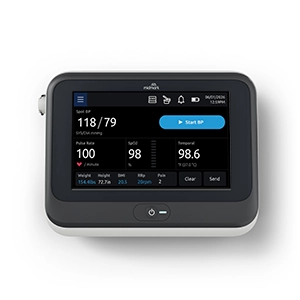
Automated oscillometric blood pressure (BP) devices detect pressure oscillations within the cuff during a BP measurement cycle. In addition to estimating systolic and diastolic pressure, device algorithms can use those oscillations to calculate a PR.
That PR is useful information, but it should be understood for what it is: a peripheral pulse-rate estimate obtained during the BP measurement period.
This creates an important workflow distinction. The measurement represents the pulses detected during a defined acquisition window rather than continuous observation of cardiac activity. Changes occurring outside that window will not be represented by that measurement.
Motion, cuff application and rhythm irregularity can also affect oscillometric measurement. For BP specifically, the American Heart Association (AHA) emphasizes use of validated devices, correct cuff selection and placement, proper patient positioning and repeated measurements when appropriate. Those fundamentals remain essential whenever BP and associated vital sign data are collected. AHA guidance on BP measurement.
An automated BP-derived PR therefore should not be interpreted as a rhythm diagnosis or as continuous cardiac monitoring.
What Pulse Oximetry Adds to the Vital Signs Workflow
A pulse oximeter uses PPG, an optical technique that detects changes in peripheral blood volume to estimate oxygen saturation and PR. Unlike a PR reported only as part of a BP cycle, a pulse oximeter can repeatedly update PR while an adequate PPG signal is being acquired.
The ability to observe PR over the measurement period can be useful during routine vital signs collection.
For example, when a single BP-derived PR appears unexpected, a PPG-derived pulse rate may provide another peripheral signal clinicians can observe while reassessing the patient. Rather than relying solely on a one-point estimate, the clinician may be able to observe whether the detected peripheral rate appears relatively stable or variable during the continuous period of measurement.
Pulse oximetry also depends on the quality of the peripheral optical signal. Poor circulation and other patient or measurement factors can affect pulse oximeter performance. The FDA advises clinicians and patients interpret pulse oximetry in the context of the overall clinical picture rather than relying on the device alone.
Technique Tip
When a PPG-derived pulse rate does not fit the patient's presentation, assess the quality and consistency of the plethysmographic (pleth) waveform, confirm sensor placement and minimize motion when possible. If available, the perfusion index (PI) can provide additional information about the strength of the pulsatile signal at the sensor site. A low or changing PI may indicate reduced peripheral pulsatile signal and should prompt closer assessment of signal quality and the patient before relying on the displayed PR.
When ECG Adds Necessary Clinical Information
Peripheral pulse measurements provide useful information about the rate, but they do not identify the underlying cardiac rhythm. When a pulse is irregular, unexpectedly fast or slow, or inconsistent with the patient’s clinical presentation, additional assessment may be needed to understand the cause.
ECG records cardiac electrical activity and provides information about both HR and rhythm. This distinction is particularly important when an irregular rhythm is suspected because a peripheral pulse signal alone cannot establish the underlying cardiac rhythm.
Current US and European atrial fibrillation (AFib) guidelines reinforce the role of ECG-based evaluation in establishing the diagnosis of AFib. The 2024 European Society of Cardiology guideline specifically distinguishes ECG documentation from non-ECG technologies such as PPG when confirming AFib for clinical management.
The 2023 ACC/AHA/ACCP/HRS guideline similarly provides an evidence-based framework for diagnosis and management of AFib.
At the point of care, an unexpectedly fast, slow or irregular peripheral rate should therefore prompt clinical reassessment. Depending on the patient's symptoms, clinical status and suspected rhythm disturbance, ECG may be appropriate to determine cardiac rate and characterize rhythm.
For patients with signs of hemodynamic instability or other acute deterioration, evaluation and escalation should follow the appropriate clinical pathway rather than relying on a peripheral PR value alone.
Clinical Application: Match the Measurement to the Question
A practical approach during routine vital signs collection is to begin with the clinical question rather than assuming that all rate measurements are equivalent.
During routine vital signs collection
Use validated devices and correct measurement techniques. For BP, ensure appropriate cuff size and placement, patient positioning and repeat measurements when indicated. A BP-device-derived PR provides an intermittent peripheral PR estimate obtained during that measurement.
When pulse oximetry is indicated
PPG-derived PR can provide repeatedly updated peripheral pulse information while adequate signal is present. Consider signal quality, sensor placement, motion and perfusion when interpreting the displayed rate.
When a rate is unexpected
Reassess the patient. Verify technique and signal quality. Repeat the measurement and compare it with other available clinical information rather than assuming the first displayed value is correct.
When the pulse is irregular or does not fit the clinical picture
Consider whether ECG assessment is warranted based on the patient's presentation and the clinical question.
When rhythm identification matters
Use ECG-based assessment. PPG and oscillometric pulse signals can identify pulse characteristics or abnormalities that warrant further evaluation, but they do not replace ECG for rhythm diagnosis.
Key Takeaways
- Heart rate and pulse rate are related but physiologically distinct. HR describes cardiac activity, while PR reflects detectable peripheral arterial pulsations.
- Automated BP devices generally derive PR from cuff oscillations during the BP measurement period. The resulting value is an intermittent peripheral estimate, not continuous cardiac monitoring.
- Pulse oximetry derives PR from a PPG signal. It can provide repeatedly updated peripheral pulse information while an adequate signal is present, but motion, perfusion and other factors can affect measurement quality.
- Neither BP-derived PR nor PPG-derived PR should be treated as a rhythm diagnosis.
- ECG provides cardiac electrical rate and rhythm information and becomes important when rhythm identification or greater diagnostic certainty is needed.
- Technique still matters. Validated devices, appropriate sensor or cuff placement, correct patient positioning and repeat measurements when indicated remain foundations of reliable vital signs collection, including pulse and heart rates.
Explore Midmark diagnostic solutions designed to support clinical workflows.
Additional Reading
- From Snapshot to Story: How Trends in Vital Signs Predict Clinical Outcomes
- The ECG Is Only as Good as the Acquisition: Why ECG Accuracy Starts with Technique
- When Every Second Counts: The Clinical Value of ECG Testing in Primary Care
- Why Automated Vital Signs Acquisition Matters in Ambulatory Care
About the Author
With a background in critical care and trauma nursing and an MBA focused on the medical device industry, I bring both clinical and business perspective to my role. My experience in the ICU and as a care flight nurse reinforced the importance of reliable equipment, standardized processes and strong clinical judgment in driving patient outcomes. As Clinical Solutions Advisor at Midmark, I partner with customers and cross-functional teams to address complex clinical needs, support product performance and strengthen clinical alignment across the product lifecycle. I am passionate about the connection between clinical accuracy, workflow and technology—ensuring healthcare professionals have both the tools and the practical insight needed to deliver high-quality care.